Brachytherapy for Cervix Cancer in AU & NZ
When radiation oncologists use radiation therapy for cervix cancer the goal is to deliver a curative dose of radiation to the tumour while sparing healthy body parts.
Radiation oncologists use brachytherapy and external beam radiation therapy to treat cervix cancer.
External beam radiation therapy is delivered from outside the body. Radiation oncologists use it to treat the cancer and areas where it has spread, such as the lymph nodes.
Brachytherapy is when doctors put radiation right in the tumour while sparing healthy body parts. Brachytherapy on its own can cure some early cervix cancers.
Most centres in Australia do high dose rate brachytherapy, which is when people get a high dose of radiation in a very short time (usually minutes) while they are under an anaesthetic.
During low dose rate brachytherapy, people get treatment over a longer time (usually several days), which means they must stay in hospital.
Both high dose and low dose rate brachytherapy are effective for treating cervix cancer.
What is the procedure for treatment?
A special team which includes a radiation oncologist, surgeon, radiation therapist , medical physicist, radiologist and anaesthetist does brachytherapy.
To prepare for brachytherapy you have an examination and blood tests. Some people also have an MRI. Doctors sometimes recommend a special diet before the procedure and an enema.
People usually have brachytherapy under a general anaesthetic, which means fasting for 6 hours before the operation. Sometimes, doctors do an anaesthetic in the back, this is called a spinal or epidural, to block pain in the lower part of the body.
People get brachytherapy in a special treatment room, and it takes 20-30 minutes.
During the process doctors insert a urinary catheter into the bladder, open the cervix and place hollow tubes, or needles, next to the cervix and tumour. These are called applicators. The doctors use an ultrasound to make sure the applicators are in the right spot and sometimes an x-ray to double check.
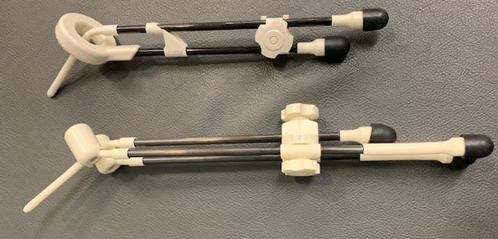
There are many different applicators, and your radiation oncologist will choose the one that will work best for you. The picture below shows 2 different types.
The radiation oncologist uses Vaseline gauze or stitches to keep the applicator in the right place.
Doctors do a scan to check the applicators are in the right spot before starting treatment. In some centres, you will be woken for the scan after the applicators are inserted. The radiation oncologist uses these images to carefully plan the treatment. After doctors have confirmed and checked they bring you in for treatment. Some centres do the planning and treatment while you’re under the general anaesthetic.
During treatment, the radioactive source moves from a shielded container through cables into the applicators. After treatment, the radioactive source goes back into the shielded container. You can’t feel the treatment.
If another treatment is needed the next day, you’ll stay in hospital overnight. Once the treatment is done, doctors take out the applicators and catheter. You may need special stockings to reduce the chance of a blood clot and can have pain relief.
For people having one treatment, it may be possible to go home the same day. Most women have 3-4 brachytherapy treatments, and these are all done under an anaesthetic.
After brachytherapy it’s important to watch for side effects such as bleeding, pain and a burning sensation when peeing. The treatment team advises people how to manage these side effects.
The treating team will schedule a follow up appointment when you leave hospital.
Potential early side effects from brachytherapy
Risks of the procedure include:
- Usual risks from an anaesthetic
- Slightly higher chance of infection
- Bleeding
- Deep vein thrombosis, which is a blood clot from lying in the same position for some hours
- Perforation, this happens if the applicator is not in the centre of the uterus but goes out through the wall. The risk of this is small due to ultrasound guidance, but if it happens the patient is usually not treated and stay in hospital overnight and take antibiotics.
- Discomfort from the applicators in place, which gets better when they are taken out.
Long term side effects post brachytherapy
- Bowel effects such as changes in bowel habits and bleeding
- Urinary issues such as bleeding in the pee
- Sexual dysfunction including pain with intercourse, dryness and narrowed vagina
Results of brachytherapy
Brachytherapy is an important part of the treatment for cervix cancer and improves outcomes.
Brachytherapy is more effective when you get it within a certain time and your treatment team schedules sessions to allow for this.