Brain Cancer Treatment in AU and NZ

Brain tumours happen when normal brain cells become abnormal and grow in an uncontrolled manner.
There are 2 types of brain tumours: primary and secondary.
Primary brain tumours
These start in the brain and rarely spread. Primary brain tumours can be benign or malignant.
Benign brain tumours are non-cancerous. They grow slowly and may take years to cause symptoms. Malignant brain tumours grow quickly. There are more than 100 different types of primary brain tumours. The most common are gliomas.
Secondary brain tumours
Secondary brain tumours are also called brain metastases. These are when cancer cells from other parts of the body spread to the brain. For example, when breast cancer cells travel from the breast through the bloodstream to the brain. Brain metastases are the most common type of brain tumours.
Radiation Therapy and Brain Cancer
The best person to talk to about radiation therapy for brain cancer is a radiation oncologist. A radiation oncologist is a specialist doctor who is part of the team that takes care of people having radiation therapy.
You can ask your doctor for a referral to a radiation oncologist to learn if radiation therapy is an option for you.
The Treatment Team
Doctors create a treatment plan for each person based on:
• the type of brain tumour
• the tumour grade
• where the brain tumour is
• the person’s health.
The type of treatment a person gets is worked out by a team of doctors and health professionals often called a Multidisciplinary Team.
A highly trained radiation oncology team takes care of people having radiation therapy. This includes radiation oncologists, radiation therapists, medical physicists and radiation oncology nurses.
Treatments for Brain Cancer
A treatment plan may include surgery, systemic therapy and radiation therapy.
Surgery
This is an important tool for diagnosing and treating brain tumours.
Surgery can include a biopsy, which is when doctors take a sample of cells from the brain to study under a microscope. Doctors use this sample to rate the tumour from low to high grade and work out the best treatment.
Surgery also includes debulking, which is when doctors remove as much of the tumour as they can. Sometimes operating is not possible if the tumour is in a part of the brain where surgery is risky.
Also, if the tumour is too large to operate or the patient is unwell the doctor will suggest other options.
Systemic therapy
This is when medicine such as chemotherapy and/or biological therapy is used to treat brain tumours. Doctors may suggest this treatment for some brain tumours after surgery.
Doctors sometimes use chemotherapy on its own or with radiation therapy. Doctors may also use systemic therapy if a brain tumour returns comes back after surgery or radiation therapy.
This treatment uses high-energy x-ray beams to kill cancer cells.
Radiation therapy depends on the type and grade of the brain tumour and if it can be removed with surgery.
Sometimes the treatment team will do radiation therapy just after surgery and in other cases it is done only if the tumour comes back.
When the treatment team does radiation therapy straight after surgery, they usually focus the radiation on the remaining tumour. If doctors completely removed the tumour and radiation therapy is recommended, the radiation is focused on the site where the tumour was taken from.
In some cases, the radiation oncologist treats a larger area such as the whole brain and spine.
What are the treatments for secondary brain tumours?
Secondary brain tumours, or brain metastases, are when cancer cells from other parts of the body spread to the brain.
Some people may have a cancer history but for others, doctors must find out where the cancer started using additional tests and imaging.
Brain imaging provides information on the number of tumours and their location.
Doctors may do extra imaging to learn if the cancer has spread to other organs, such as the liver, lungs or bones.
Treatment depends on:
- where the cancer started
- if other organs are affected
- how many metastases are in the brain
- where the metastases are located and their size
- a person’s overall health.
Treatments for brain metastases include surgery and stereotactic radiosurgery.
Types of Radiation Therapy used In Brain Cancer
External Beam Radiation Therapy (EBRT)
This is the most common type of radiation therapy radiation oncologists use for brain tumours.
Radiation oncologists often use Volumetric Arc Therapy (VMAT) which is an advanced types of external beam radiation therapy, to carefully deliver radiation to the areas that need to be treated.
This advanced technique allows the radiation oncologist to target the radiation on the cancer while limiting radiation to healthy parts of the body.
People get EBRT once a day, 5 days a week from Monday to Friday. This usually lasts for 1-6.5 weeks.
People often wear a plastic mask during treatment to reduce movement and ensure the radiation is targeted on the cancer.
Stereotactic Radiosurgery (SRS)
This is when the radio oncologist delivers one high dose of targeted radiation to the tumour.
SRS may be an option for secondary brain cancer if there aren’t many brain metastases and there is no cancer outside the brain, or the cancer outside the brain is well managed.
If there are many brain metastases and/or the cancer outside the brain is not well managed radiation oncologists may do radiation therapy on the whole brain.
Whole brain radiation therapy is 1 treatment a day, 5 days a week, over 1-2 weeks.
The treatment team may also use whole brain radiation therapy after surgery or stereotactic radiosurgery to reduce the risk of brain metastases coming back.
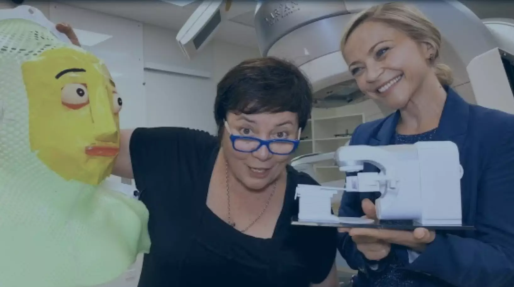
Immobilisation Mask
People wear a mask during radiation treatment to keep their head and neck still. The mask ensures the radiation beams target the cancer accurately while minimising exposure to healthy body parts nearby.
A new mask is made for each person and is secured to the treatment table. The mask helps the doctor position the radiation accurately on the cancer being treated.
In this video, Targeting Cancer ambassador Julie McCrossin explains why people with head and neck cancer need to wear protective masks during treatment.
Why We Wear Masks For Head and Neck Cancer Radiation Therapy Treatment
In this video, Julie and the radiation oncology team explain how an immobilisation mask is made.

Targeting Cancer Julie’s Story- The Making of the Immobilisation Mask
General Information About Side Effects of Radiation Therapy
Radiation therapy is more effective with fewer side effects than ever before.
Recent advances mean radiation oncologists can effectively treat the cancer while getting less radiation on healthy body parts. This means much fewer side effects.
Side effects from radiation therapy vary between people, even for those having the same treatment.
While some people feel no side effects, some feel mild side effects, such as tiredness or skin redness during and/or just after treatment. These usually get better within a few weeks.
The treatment team will offer advice and medicine to help with side effects.
Serious side effects that start later (months to years after the radiation therapy) are rare.
Before starting treatment, your radiation oncologist will talk to you about side effects and answer your questions.
The side effects of radiation treatment can be split into 2 groups:
- Early side effects which occur during and shortly after radiation treatment.
- Late side effects which can occur months to years after radiation treatment.
For more information, go to the Potential Side Effects page.
Early Side Effects of Radiation Therapy for Brain Cancer
Early side effects of radiation therapy may include:
Fatigue: This is very common in the second half of treatment and varies between people. Tiredness may continue for several weeks after treatment.
Other side effects of radiation treatment may come from body parts close to where the radiation is targeted. These often start during treatment, worsen and reach their peak in the 7-10 days following treatment
Skin reddening and irritation: The scalp may get red, dry and itchy.
Hair loss: People may lose some or all their hair. This can be short or long term, depending on amount of radiation used.
Swelling in the brain: Radiation therapy can cause swelling around the brain tumour. This may cause headaches, nausea, vomiting and/or drowsiness. The swelling can also worsen existing conditions in the short term.
Ringing in the ears and hearing loss: These are very rare side effects.
Seizures: This is uncommon but can cause changes in behaviour and movement.
What can reduce early side effects?
- Resting as needed helps with tiredness.
- Creams can be used on the scalp for red skin and irritation.
- Wigs can be used for hair loss.
- If headaches, nausea and/or vomiting occur during treatment, medicine that reduces swelling around the tumour can help.
- Pain and anti-nausea medicine can help.
Early delayed side effects
Somnolence syndrome is a rare side effect that can occur 1-6 months after radiation treatment. Signs include sleepiness, irritability, headaches, nausea, vomiting and loss of appetite. These things usually get better after a few weeks and corticosteroid medicine may help.
Late Side Effects of Radiation Therapy for Brain Cancer
Late side effects can happen a few months to a few years after treatment. These side effects may never occur, occur once, continue over time, or come and go.
They are rarer than early side effects.
The chances of late side effects depends on how much radiation is used and how close the tumour is to other parts of your body.
Late side effects of radiation treatment may include:
Neurocognitive changes: These change the way people think, learn and remember. These changes can happen after surgery and chemotherapy as well.
Less common late side effects of radiation therapy may come from body parts close to where the radiation is targeted.
Eye and optic nerves: Radiation can cause the lens of the eye to become cloudy. This can cause painless vision loss years later. Changes to the retina may also cause loss of vision. Radiation to the optic nerves sometimes causes total loss of sight.
Ears: Hearing loss and ringing in the ear can develop in the years after treatment and are usually lasting.
Pituitary gland: Radiation may cause the pituitary gland to become underactive. Your doctor may do blood tests after treatment to keep an eye on this.
Brain necrosis: This rare effect can occur 1-3 years after treatment with high doses of radiation. The symptoms vary and it may require surgical treatment.
Radiation induced cancers: Cancers caused by radiation therapy are a very rare side effect.
What can treat late side effects
- Doctors can check sight changes to work out the cause. If radiation causes a cataract, doctors can fix this with a small operation. Radiation damage to the retina or optic nerve may remain.
- Hearing aids can help with hearing loss.
- Hormone replacement can help an underactive pituitary gland.
- Medicine can help neurocognitive changes.
Your radiation oncologist will explain the types of late side effects that could happen and how you can manage them.
Other Useful Resources for Brain Cancer
Find additional information about cancer types, research groups, and support groups.
Radiation Oncologist
The best person to talk to is a radiation oncologist. You can ask your doctor for a referral to find out if radiation treatment is right for you.
GPs and Health Professionals
Information for any health professional involved in a patient's cancer care with a particular focus on primary care providers.
Talking to Your Doctor
Your GP or other doctors in the cancer team can organise a referral to a radiation oncologist.
Treatment Centres
Search and find your closest Radiation Oncology Treatment Centre.